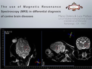
THE USE OF MAGNETIC RESONANCE SPECTROSCOPY IN THE DIFFERENTIATION OF BRAIN DISEASES
Single-voxel proton Magnetic Resonance Spectroscopy STEAM sequence can be used to investigate, non invasively, a wide range of brain diseases. At present several studies have been conducted on human patients to assess MRS reliability in primary vs. metastatic brain tumors differentiation; further papers tested MRS ability in differentiation of histological tumor types or simply, to differentiate neoplastic from non- neoplastic. The aim of this study was to investigate MRS spectra of canine single-lesion brain diseases of inflammatory, neoplastic or metabolic origin (hepatic encephalopathy).
From 2008 to 2014 a total amount of 578 patients underwent brain MRI and MRS examination upon informed consent agreement from the owners.
For this retrospective investigation MRI and MRS of patients with intracranial lesions were reviewed and patient were grouped into inflammatory, neoplastic and hepatic encephalopathy (HE). The admission criteria were, respectively for the inflammatory group all the patients that showed multiple or single well-defined lesions at MRI scan and at cerebral Diffusion Weighted Imaging (DWI) a suggestive of inflammation disease and furthermore from this selection we included only patients with pleocytosis and high total protein concentration on CSF (total cell count >5 cells/!L and total protein concentration > 30 mg/dL). For the neoplastic group all the patients with centrally or peripherally strong enhancing mass lesions with surrounding discrete edema, according to MRI findings reported in veterinary papers. For the metabolic group all the patients affected from hepatic encephalopathy diagnosed by clinical examination, blood cell count, biochemical panel, AmmoniaCheck (Menarini) determination and optional hystopatology of the liver and, finally, a large group of patients who had normal spectroscopy.
MRS was performed twice on lesion larger, one using a short TR35 and the other using a long TR144. The VOI 1x1x1cm was defined in the parieto-occipital region or where there was a greater hyperintensity. The metabolites studied were the compound of Glutamine-glutamate (Glx), the compound of N-acetylaspartate and N- acetyl aspartyl glutamate (NAA), the Choline derivatives (Cho), and the Myo-Inositol (My). The NAA/ Cr, Cho/Cr and My/Cr ratios were analyzed as the presence of lipids and lactate.
Patient selected were 578: 112 with inflammatory brain diseases, 144 with neoplastic lesions, 22 with hepatic encephalopathy. Spectra obtained by single-voxel MRS examination were divided for each category.
MRS spectra from the inflammatory lesion affected dogs showed increases of Cho (mean Cho/Cr ratio: 2.23), reductions of NAA (mean NAA/Cr ratio: 1.15) and the presence of the lipid peak were found in 46/112 cases. The mean values and their standard deviations were: Glx/Cr 0,26 std. 0,2; Cho/Cr 2.23 std. 0,9; My/ Cr 0,54 std. 0,1; NAA/Cr 1,35 std. 0,59. MRS spectra from neoplastic patients were characterized for high values of Cho and low or absent NAA. Presence of lipids and lactate were founded in 31/144 lesions. Mean values and relatives standard deviations are reported: Glx/Cr 0,22 std. 0,32; Cho/Cr 3,27 std. 0,69; My/Cr 0,54 std. 0,11; NAA/Cr 1,14 std. 1,03.
The HE grouped patient displayed decreased values of My, Cho and NAA as well as increased values of Glx. Mean values and connected standard deviations were: Glx/ Cr 0,40 std. 013; Cho/Cr 1,14 std. 0,34; My/ Cr 0,15 std. 0,08; NAA/Cr 1,02 std. 0,35. The group considered normal was composed of 300 patients showed values of Cho, Cr and NAA widely similar to those obtained in human medicine and in particular: Glx/Cr 0,21 std. 0,08; Cho/Cr 1,58 std. 0,22; My/Cr 0,54 std. 0,11; NAA/Cr 1,92 std. 0,23. In vivo single-voxel proton MRS provides a sensible and non-invasive characterization of canine brain lesions. Reliable patterns of differentiation based on NAA/Cr and Cho/Cr values and on presence of lipids can be used in differential diagnosis of inflammatory, neoplastic and metabolic diseases.
The images produced by spectroscopy have also proved useful in the planning of radiotherapy treatment plans for brain and of course for the assessment of the potential damage caused by radiation therapy in the follow-up of patients who underwent a brain radiosurgery.
